Neutrophil recruitment and activation are differentially dependent on MyD88/TRIF and MAVS signaling during RSV infection
Neutrophil recruitment and activation are differentially dependent on MyD88/TRIF and MAVS signaling during RSV infection"
- Select a language for the TTS:
- UK English Female
- UK English Male
- US English Female
- US English Male
- Australian Female
- Australian Male
- Language selected: (auto detect) - EN
Play all audios:
Respiratory syncytial virus (RSV) is a leading cause of severe lower respiratory tract infections, especially in infants. Lung neutrophilia is a hallmark of RSV disease but the mechanism by
which neutrophils are recruited and activated is unclear. Here, we investigate the innate immune signaling pathways underlying neutrophil recruitment and activation in RSV-infected mice. We
show that MyD88/TRIF signaling is essential for lung neutrophil recruitment while MAVS signaling, leading to type I IFN production, is necessary for neutrophil activation. Consistent with
that notion, administration of type I IFNs to the lungs of RSV-infected Mavs−/− mice partially activates lung neutrophils recruited via the MyD88/TRIF pathway. Conversely, lack of neutrophil
recruitment to the lungs of RSV-infected Myd88/Trif−/− mice can be corrected by administration of chemoattractants and those neutrophils become fully activated. Interestingly, Myd88/Trif−/−
mice did not have increased lung viral loads during RSV infection, suggesting that neutrophils are dispensable for viral control. Thus, two distinct pathogen sensing pathways collaborate
for neutrophil recruitment and full activation during RSV infection.
Neutrophils are classically considered as among the first immune cells to respond to both infection and injury. Historically, neutrophils were considered as non-specific effector cells but
it is becoming increasingly clear that they can respond differentially to harmful stimuli.1 Pattern recognition receptor (PRR) signaling induces innate immune responses including the
generation of chemotactic gradients along which neutrophils are recruited into affected tissues.2,3 Following recruitment, additional signals are required to trigger neutrophil activation
and degranulation. During activation, neutrophils can secrete hundreds of pre-stored effector proteins and proteolytic enzymes including matrix metalloproteinases (MMPs), myeloperoxidase
(MPO), and neutrophil elastase (NE).4 Neutrophils can also drive inflammation by generating reactive oxygen species (ROS), as well as by undergoing a unique form of cell death whereby they
secrete nuclear DNA in the form of neutrophil extracellular traps (NETs).4,5,6 Whilst neutrophils are important as immediate antimicrobial responders, their recruitment and activation must
be tightly regulated to minimalize pathology caused by bystander effects on host tissues.7,8 This is particularly relevant in the lung where the disruption of delicate alveolar structures
can rapidly have life-threatening consequences if gas exchange is compromised. The molecular mechanisms regulating neutrophil recruitment and activation in response to bacterial and fungal
respiratory infections have been well characterized.9 However, considerably less is known about the regulation of neutrophil action during respiratory viral infections.10,11,12
Respiratory syncytial virus (RSV) is the greatest cause of infant hospitalizations in the developed world.13,14 More than 90% of children encounter RSV within their first 24 months and a
minority develops severe disease; 1–3% will require hospitalization.15 In addition to acute disease, RSV-induced bronchiolitis and viral pneumonia in infancy are associated with
debilitating, long-term respiratory disorders, such as recurrent wheeze16 and asthma.17 Although it is not well understood why some infants are asymptomatic while other infants develop
severe disease, host immune factors have been implicated in disease severity.18,19 During RSV infection, neutrophils are recruited to the lungs of both mouse and man.20,21,22 Clinical
studies suggest that neutrophils contribute to immune pathology during disease20,22,23 and they are the most abundant cell type in the bronchoalveolar lavage (BAL) of infants with
RSV-induced bronchiolitis.20,22 Neutrophil activation during RSV infection has been more difficult to assess clinically than neutrophil recruitment. However, elevated NE concentrations have
been observed in both the serum and nasal lavage of children with confirmed RSV infection.23 Together, these studies suggest that a hyperactive or dysregulated neutrophil response to RSV may
underlie disease severity and indicate the importance of studying the signaling pathways that regulate neutrophil recruitment and activation in the infected lung.
Innate immune receptors on epithelial cells and alveolar macrophages (AMs) initiate the immune response to RSV within the respiratory tract.24,25 The plasma membrane expressed Toll-like
receptors (TLRs), TLR2/TLR6 and TLR4, and the endosomally-expressed TLR3 and TLR7, have all been implicated in RSV detection.25,26 RSV is also detected in the cytosol by retinoic
acid-inducible gene I (RIG-I)-like receptors (RLRs): RIG-I and melanoma differentiation–associated protein 5 (MDA5).27,28,29 RLR signaling occurs through the adaptor protein mitochondrial
antiviral signaling protein (MAVS)30,31 while TLRs signal via the adaptor proteins MyD88 and/or TRIF.32 Signaling via MAVS, MyD88 and TRIF induces the expression of type I and type III
interferons (IFNs), as well as other pro-inflammatory cytokines and chemokines.33 Type I IFNs bind to the interferon-α/β receptor (IFNAR) expressed on all nucleated cells and induce the
expression of hundreds of IFN-stimulated genes (ISGs).34 ISG protein products have direct antiviral properties and can also promote the recruitment and activation of other components of the
immune response, including antiviral monocytes.21,27
In this study, we investigated the necessity of MAVS and MyD88/TRIF dependent signaling pathways to lung neutrophil recruitment and activation during RSV infection in vivo. Mavs−/− mice,
unable to signal via MDA-5 or RIG-I, and Myd88/Trif−/− mice, unable to signal via TLRs (and the IL-1 receptor (IL-1R) and IL-18 receptor (IL-18R)) were infected with RSV. We found that
neutrophil recruitment to the lung was dependent on MyD88/TRIF signaling but mostly independent of MAVS signaling. The neutrophil chemoattractant CXCL1 was shown to be mainly produced by
non-epithelial, non-endothelial (CD45−, Epcam−, CD31−) lung cells. However, neutrophil activation in the lung was dependent on the type I IFN-driven, pro-inflammatory lung environment
induced by MAVS signaling. Restoring the pro-inflammatory environment in the lungs of Mavs−/− mice using recombinant IFN-α (rIFN-α) was sufficient to partially activate neutrophils during
RSV infection. Conversely, neutrophils recruited into the lungs of Myd88/Trif−/− mice using recombinant CXCL1 (rCXCL1) became fully activated during RSV infection. This study sheds light on
the molecular mechanisms that drive neutrophil recruitment and activation in the lungs during RSV infection and demonstrates how two distinct PRR signaling pathways collaborate to drive
neutrophil responses to virus infection.
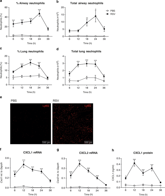
Neutrophils are recruited to the lungs during RSV infection in both mice and man.20,23,27 To establish the exact timing of neutrophil recruitment to the airways and lungs in the C57BL/6
mouse model, mice were intranasally (i.n.) infected with RSV. The frequencies and total numbers of neutrophils were quantified over time in the airways, by sampling the BAL, and in the lungs
using flow cytometry (Fig. 1a–d). Neutrophils were defined as CD45+, CD3−, CD19−, MHC II−, Ly6G+ cells (gating strategy shown in Supplementary Fig. 1). Neutrophil recruitment to the airways
and to the lung peaked at 18–24 h post infection (p.i.) and was not induced by mock (PBS) infection (Fig. 1a–d). Fluorescence microscopy of lung sections demonstrated that Ly6G+ cells were
evenly distributed throughout the lungs during RSV infection at 24 h p.i. (Fig. 1e). CD64+ inflammatory monocyte recruitment to the lung also occurred early following RSV infection
(Supplementary Fig. 2a, b), with their recruitment peaking after lung neutrophilia has resolved at 48 h p.i.,27 but, in contrast, RSV infection did not have much of an impact on the number
of AMs in the airways at any of the early timepoints (Supplementary Fig. 2c). Expression of the neutrophil chemoattractants Cxcl1 and Cxcl2 peaked prior to neutrophil influx at 12 h p.i. and
this was confirmed at the protein level for CXCL1 (Fig. 1f–h).
Neutrophil recruitment to the lung during RSV infection in vivo. Wt mice were intranasally mock (PBS) or RSV infected. a–d Frequencies and total numbers of CD45+, CD3−, CD19−, MHC-II−, Ly6G+
neutrophils were quantified in the airways and lung using flow cytometry at the indicated time-points (Supplementary Fig. 1a for gating strategy). e Images show representative
immunostaining of Ly6G+ neutrophils (red) in lung cryosections at 24 h p.i. from three mice per group. f, g Cxcl1 and Cxcl2 was quantified relative to Gapdh by RT-qPCR from RNA isolated from
lung tissue. h CXCL1 was detected in the BAL fluid by ELISA. Data are presented as the mean ± SEM of 3–5 (PBS), or 8–13 (RSV) individual mice, pooled from at least two independent
experiments. Statistical significance of differences between mock and RSV infection was determined by one-way ANOVA with Tukey’s post hoc test. *P ≤ 0.05, **P ≤ 0.01, ***P ≤ 0.001
Next, we assessed the activation status of neutrophils recruited to the airways and lung during RSV infection (Fig. 2). Neutrophil activation status was initially determined by the detection
of the proteolytic enzymes MMP-9, MPO and NE, all of which are pre-stored in cytoplasmic granules and released during neutrophil degranulation.35 Whilst neutrophil recruitment peaked at
18–24 h p.i., the secretion of MMP-9, MPO and NE into the airways, as detected in the BAL fluid, peaked at 24 h p.i. (Fig. 2a). Neutrophils were confirmed to be the major cellular source of
MMP-9 and NE in BAL as these mediators were not detected after anti-Ly6G depletion (Fig. 2b, c). In contrast, neutrophil depletion with anti-Ly6G reduced but did not entirely abrogate MPO
levels in BAL suggesting that there could be additional cellular sources of MPO in the airways after RSV infection (Fig. 2c).
Neutrophil activation in the lung during RSV infection in vivo. Wt mice were intranasally mock (PBS) or RSV infected. a MMP-9, MPO and NE were detected in the BAL fluid by ELISA. b, c Mice
were given 200 μg i.n. and 500 μg i.p. α-Ly6G or isotype control antibody 1 day before infection. After 18 h p.i., b frequencies and total number of neutrophils in the airways were
quantified (the frequencies of neutrophils were determined by differential counting of >300 cells on H&E stained cytospin slides) or c MMP-9, MPO and NE were detected in the BAL fluid by
ELISA. d Representative histograms of CD64 expression on CD45+, CD3−, CD19−, MHC-II−, Ly6G+ neutrophils in the lung 18 h p.i. (Supplementary Fig. 1a for gating strategy) e Frequencies and
total numbers of CD64+ lung neutrophils (CD45+, CD3−, CD19−, MHC-II−, Ly6G+), quantified using flow cytometry. Data from the time course are presented as the mean ± SEM of 3–5 (PBS) or 8–13
(RSV) individual mice pooled from two independent experiments. Data from the depletion experiments are presented as the mean ± SEM from 4–5 (PBS) or 7–8 (RSV) individual mice, pooled from
two independent experiments. Statistical significance of differences were determined by one-way ANOVA with Tukey’s post hoc test. *P ≤ 0.05, **P ≤ 0.01, ***P ≤ 0.001
To further assess neutrophil activation status during RSV infection, we quantified the cell surface expression of a variety of markers associated with neutrophil activation in other
inflammatory contexts on lung, airway and peripheral blood neutrophils (Supplementary Fig. 3). Upregulation of CD64, CD69 and CD11b on the cell surface of neutrophils has previously been
associated with neutrophil activation,36,37,38 as has the downregulation of CD62L and CD182.39,40 During RSV infection, we found that CD69 expression remained low on neutrophils in all
tissues, regardless of infection status. CD11b was upregulated upon migration into the airways and lungs, but this was also seen in the absence of an active RSV infection. Likewise, CD62L
and CD182 were downregulated on airway and lung neutrophils relative to blood neutrophils, but not specifically in response to RSV infection (Supplementary Fig. 3). Of the activation markers
tested, CD64 was the most specific marker of neutrophil activation in the lungs (Fig. 2d, e and Supplementary Fig. 3). CD64 expression levels were low on blood neutrophils and high on lung
and airway neutrophil during RSV infection (Supplementary Fig. 3), peaking at 18–24 h p.i. (Fig. 2e). We confirmed that mock infection did not induce the secretion of MMP-9, MPO or NE into
the airways or the upregulation of cell surface activation marker CD64 at any time point (Fig. 2). We also confirmed that live virus, and not any mediator in the virus preparation, was
necessary for neutrophil recruitment and activation as ultra-violet inactivated RSV (UV RSV) did not recruit neutrophils, induce MMP-9, MPO or NE secretion, or drive CD64 cell surface
upregulation on lung neutrophils (Supplementary Fig. 4). Also, HEp-2 cell supernatant did not induce neutrophil recruitment (data not shown). Together, these data confirm that neutrophils
are a major cell type recruited to the lungs early during RSV infection in the murine model and demonstrate that RSV infection induces neutrophil activation in the lung.
RSV is recognized by both RLRs and TLRs, which signal via the cytoplasmic adaptor proteins MAVS and MyD88/TRIF, respectively.25,26 To investigate the lack of redundancy of each of these two
distinct signaling pathways in neutrophil recruitment, Mavs−/− and Myd88/Trif−/− mice were infected with RSV and airway neutrophils were quantified at 18 h p.i. (Fig. 3a). After RSV
infection, neutrophils composed 40% of the airway cells in wild-type (wt) mice and 25% of the airway cells in Mavs−/− mice. No neutrophils were recruited during RSV infection in
Myd88/Trif−/− mice (Fig. 3a). These data indicate that MyD88/TRIF signaling is essential for neutrophil recruitment to the lungs during RSV infection as has previously been implied in
Myd88−/− mice.41 As reported,27,28,29 Mavs−/− mice had a higher viral load than wt mice at 18 h p.i. and at day 4 p.i. (the peak of viral load in the mouse model21). In contrast,
Myd88/Trif−/− mice had a similar viral load to wt mice at both timepoints measured (Supplementary Fig. 5).
Neutrophil recruitment to the lung and CXCL1 gene induction in stromal cells are dependent on MyD88/TRIF signaling during RSV infection. Wt, Mavs−/− and Myd88/Trif−/− mice were intranasally
mock (PBS) or RSV infected. a, b Percentage and total number of neutrophils in the airways 18 h p.i. The frequency of neutrophils was determined by differential cell counting of >300 cells
on H&E stained cytospin slides. Data are presented as the mean ± SEM from 3–5 (PBS) or 8 (RSV) individual mice (represented as individual dots) pooled from two independent experiments. b–d
Wt and Myd88/Trif−/− mice were mock (PBS) or RSV infected for 12 h. RSV L gene copies, Ifna5 and Cxcl1 were quantified relative to Gapdh by RT-qPCR from RNA isolated from fluorescence
activated cell sorted AMs (CD45+, CD11c+), ATII epithelial cells (CD45−, EpCAM+, CD31−), endothelial cells (CD45-, EpCAM-, CD31+) and other stromal cells (CD45−, EpCAM−, CD31−; Supplementary
Fig. 1b for gating strategy) Data are presented as mean ± SEM of 1–3 sorts per group (represented as individual dots), with 3–6 mice pooled per sort. In a statistical significance of
differences was determined by two-way ANOVA with Tukey’s post hoc test. ***P ≤ 0.001 and in d statistical significance of differences between wt sorted cell populations were determined by
two-way ANOVA with Bonferroni’s post hoc test. **P ≤ 0.01
To further investigate the mechanism driving neutrophil recruitment to the lung during RSV infection, we assessed the source of one of the neutrophil chemoattractants, CXCL1. Wt and
Myd88/Trif−/− mice were mock (PBS) or RSV infected and resident AMs, endothelial cells, ATII epithelial cells and CD45−, EpCAM− CD31− cells (other stromal cells) were sorted at 12 h p.i.
(Supp. Fig. 1b for FACS gating strategy) and compared to a presorted sample (total lung). RSV L gene transcripts were detected in all cell populations in RSV-infected mice (Fig. 3b). Ifna5
expression was detected in the total lung sample (presort) and in sorted AMs (Fig. 3c) from both wt and Myd88/Trif−/− mice, confirming previous findings.24,27 Interestingly, Cxcl1 mRNA was
significantly enriched in the CD45-, EpCAM-, CD31- cell population (other stromal cells) from wt mice, suggesting that non-epithelial, non-endothelial cells are the major source of CXCL1
during RSV infection (Fig. 3d). Cxcl1 mRNA was not detected in any lung cell populations from RSV-infected Myd88/Trif−/− mice (Fig. 3d). These data suggest that MyD88/TRIF signaling is
crucial for recruitment of neutrophils into the lungs during RSV infection and that signaling via MyD88/TRIF is necessary for inducing Cxcl1 and, possibly, other neutrophil chemoattractants
in the lung stromal cell compartment.
We next investigated how the MAVS and MyD88/TRIF dependent PRR signaling pathways regulate neutrophil activation in the lung during RSV infection (Fig. 4a–c). MMP-9, MPO and NE were measured
in the airways of wt, Mavs−/− and Myd88/Trif−/− mice at 18 h p.i. (Fig. 4a). Consistent with their inability to recruit neutrophils to the lung, Myd88/Trif−/− mice did not display increased
levels of MMP-9, MPO or NE in the airways after RSV infection (Fig. 4a). Notably Mavs−/− mice, which did recruit neutrophils during RSV infection, also did not show elevated MMP-9, MPO or
NE in BAL during RSV infection (Fig. 4a). Furthermore, lung neutrophils from RSV-infected Mavs−/− mice lacked cell surface CD64 expression (Fig. 4b, c). In contrast, the very few lung
neutrophils found in RSV infected Myd88/Trif−/− mice (that have functional MAVS signaling) did upregulate CD64 normally (Supplementary Fig. 6). These data suggest that MAVS signaling is
required for neutrophil activation in the lung during RSV infection, at least as measured by CD64 upregulation and the secretion of some granule contents.
Neutrophil activation is dependent on MAVS signaling during RSV infection. Wt, Mavs−/− and Myd88/Trif−/− mice were intranasally mock (PBS) or RSV infected for 18 h. a MMP-9, MPO and NE were
detected in the BAL fluid by ELISA. b Representative histograms of CD64 expression on CD45+, Ly6G+ neutrophils in the lung. c Frequencies of CD64+ expression on CD45+, Ly6G+ lung
neutrophils, quantified using flow cytometry. d IFN-α and IL-6 were detected in the BAL fluid by ELISA. Data are presented as the mean ± SEM from 3–5 (PBS) or 8 (RSV) individual mice
(represented as individual dots) pooled from two independent experiments. Statistical significance of differences was determined by two-way ANOVA with Tukey’s post hoc test. *P ≤ 0.05, **P ≤
0.01, ***P ≤ 0.001
Signaling via MAVS is essential for the production of type I IFNs and lung inflammation during RSV infection.27 We confirmed that IFN-α and IL-6 were not present in the airways of Mavs−/−
mice at 18 h p.i. (Fig. 4d). However, Myd88/Trif−/− mice had comparable levels of IFN-α and IL-6 in the BAL to wt mice (Fig. 4d). Therefore, although signaling via MyD88/TRIF is required for
neutrophil recruitment during RSV infection, it is not required for the overall establishment of a cytokine-driven, pro-inflammatory lung environment as assessed by IFN-α and IL-6 levels.
These observations lead to the hypothesis that, during RSV infection, pro-inflammatory factors induced downstream of MAVS signaling activate lung neutrophils after they are recruited into
the lungs as a downstream consequence of MyD88/TRIF signaling.
We tested this hypothesis firstly by restoring the pro-inflammatory environment in the lungs of Mavs−/− mice during RSV infection by intranasal administration of rIFN-α, which induces
pro-inflammatory cytokines and monocyte recruitment in the lungs of wt mice.21 We confirmed that this also works in Mavs−/− mice (Fig. 5a, b) and in mock infected wt mice (Supplementary Fig.
7a) without impacting neutrophil or AM numbers (Fig. 5c, d and Supplementary Fig. 7b). Consistent with induction of pro-inflammatory mediators, rIFN-α but not BSA (a protein control for
rIFN-α) was sufficient to cause upregulation of CD64 on lung neutrophils present at baseline in wt mice (Supplementary Fig. 6c). Likewise, rIFN-α was sufficient to drive upregulation of CD64
in RSV-infected Mavs−/− mice to levels comparable to those in RSV-infected wt mice (Fig. 5e, f). This was accompanied by higher levels of MPO in the BAL (Fig. 5g) and a trend for increased
levels of airway MMP-9 and NE, which did not reach statistical significance (Fig. 5g). Thus, rIFN-α is sufficient to drive an inflammatory environment in the lung that results, at least
partially, in neutrophil activation in RSV-infected Mavs−/− mice.
rIFN-α restores neutrophil activation in the lung of MAVS deficient mice during RSV infection. Wt and Mavs−/− mice were mock (PBS) or RSV infected for 24 h. PBS or 1 μg rIFN-α was
administered i.n. 6 h p.i. a mRNA was quantified by RT-qPCR from RNA isolated from the lung tissue. Expression levels of Il6 and Csf2 were quantified relative to Gapdh. Absolute levels of
IFNg and Tnfa were quantified using a plasmid standard. b Total number of lung monocytes as quantified by flow cytometry (Supplementary Fig. 1a for gating strategy). c Total number of airway
AMs as quantified by flow cytometry. d Total number of airway and lung CD45+, CD3−, CD19−, MHC-II−, Ly6G+ neutrophils, as quantified by flow cytometry. e Representative histograms of CD64
expression on CD45+, CD3−, CD19−, MHC II−, Ly6G+ neutrophils in the lung. f Frequency of CD64+ lung neutrophils, as quantified by flow cytometry. g MMP-9, MPO and NE were detected in the BAL
fluid by ELISA. Data are presented as mean ± SEM of 8–17 mice (represented as individual dots) from each group, pooled from 3–4 independent experiments. Statistical significance of
differences was determined by one-way ANOVA with Tukey’s post hoc test. Only the statistical significances between Mavs−/− RSV/PBS and RSV/IFN-α are shown. *P ≤ 0.05, **P ≤ 0.01, ***P ≤
0.001
Secondly, we used Myd88/Trif−/− mice to test our hypothesis that factors induced downstream of MAVS signaling activate lung neutrophils during RSV infection. Myd88/Trif−/− mice produce type
I IFNs in response to RSV as they have intact MAVS signaling and therefore have a highly pro-inflammatory lung environment early during RSV infection (Fig. 4). We postulated that
Myd88/Trif−/− mice do produce the neutrophil activating factors in the lung in response to RSV, despite neutrophil recruitment being abrogated. Indeed, as mentioned, the few neutrophils that
can be found in RSV infected Myd88/Trif−/− mice upregulated the activation marker CD64 (Supplementary Fig. 6). However, to further test this hypothesis, Myd88/Trif−/− mice were treated with
rCXCL1 i.n. 6 h p.i. to recruit neutrophils into the lung after RSV infection (Fig. 6). Neutrophil responses were evaluated at 18 h p.i. Treatment of Myd88/Trif−/− mice with rCXCL1 during
RSV infection did not influence the number of airway AMs or lung monocytes as compared to mock treatment (Fig. 6a, b).
Neutrophil activation is not dependent on MyD88/TRIF signaling in the lung during RSV infection. Wt and Myd88/Trif−/− mice were mock (PBS) or RSV infected for 18 h. PBS or 10 μg rCXCL1 was
administered i.n. 6 h p.i. a Total number of airway AMs as quantified by flow cytometry. b Total number of lung monocytes as quantified by flow cytometry. c Total numbers of airway and lung
CD45+, CD3−, CD19−, MHC-II−, Ly6G+ neutrophils as quantified by flow cytometry. d Total number of CD64+ lung neutrophils, as quantified by flow cytometry. e MMP-9, MPO and NE were detected
in the BAL fluid by ELISA. Data are presented as mean ± SEM of 6–12 mice from each group (represented as individual dots), pooled from 2–3 independent experiments. Statistical significance
of differences was determined by one-way ANOVA with Tukey’s post hoc test. Only the statistical significances between groups of MyD88/Trif−/− mice are shown. *P ≤ 0.05, **P ≤ 0.01, ***P ≤
0.001
As previously observed, Myd88/Trif−/− mice did not recruit neutrophils to the airways or lungs 18 h p.i. (Fig. 6c) but treatment with 10 μg rCXCL1 was sufficient to recruit neutrophils (Fig.
6c). There was no difference in neutrophil recruitment between mock infected or RSV infected Myd88/Trif−/− mice treated with rCXCL1 (Fig. 6c). Lung neutrophils recruited into RSV-infected
Myd88/Trif−/− mice displayed an activated phenotype as measured by the upregulation of cell surface CD64, which was comparable to that observed on neutrophils during RSV infection of wt mice
(Fig. 6d). CXCL1 treatment in the absence of RSV did not cause upregulation of CD64 on lung neutrophils in Myd88/Trif−/− mice (Fig. 6d), confirming that the MAVS-dependent inflammatory
environment is required for full neutrophil activation. On its own, rCXCL1 did induce the accumulation of some MMP-9, MPO and NE in the BAL of Myd88/Trif−/− mice (Fig. 6e), but this was
significantly increased during RSV infection. Together, these data demonstrate that the factors which activate lung neutrophils during RSV infection are present in the lungs of Myd88/Trif−/−
mice despite the fact that these mice cannot recruit neutrophils to the lung during the infection.
Neutrophils are one of the first cell types to be recruited to the lung during RSV infection21 and lung neutrophilia is a hallmark of severe RSV infection in infants.20,22,23 Here, innate
immune signaling deficient Mavs−/− and Myd88/Trif−/− mice were used to understand which RSV sensing pathways are required for the early recruitment and activation of lung neutrophils during
RSV infection. Interestingly, we found that both MyD88/TRIF and MAVS signaling are required but that they control distinct aspects of the neutrophilic response (Fig. 7). Indeed, while
MyD88/TRIF signaling was necessary for neutrophils to infiltrate the lung, it was dispensable for their activation whereas the opposite was true for the MAVS pathway. To our knowledge, this
is the first study to demonstrate that distinct innate immune signaling pathways synergize to drive lung neutrophilia and neutrophil activation during a respiratory virus infection.
Schematic to illustrate the mechanism by which neutrophils are recruited and activated in the lung during RSV infection. RSV enters the airways (1) and interacts either directly or
indirectly with stromal cells to induce the production of CXCL1 via MYD88/TRIF signaling (2). This drives the recruitment of neutrophils into the lungs from the vasculature (3). Meanwhile,
AMs produce type I IFNs in response to RSV via signaling through MAVS (4). Type I IFNs drive the induction of other pro-inflammatory mediators, such as TNF-α and IL-6, and chemokines such as
CCL2 by multiple other cell types including DCs and monocytes (5). When neutrophils encounter this pro-inflammatory environment, they upregulate cell surface CD64 and become activated to
secrete MMP-9, MPO, and NE (6). The illustration was created using BioRender
Neutrophils are classically considered rapid responders and, consistent with that, early neutrophil recruitment was detected in the lung post RSV infection. To assess the activation status
of lung neutrophils, the cell surface expression of known activation markers was investigated by flow cytometry. This provides a sensitive readout of neutrophil activation as the expression
level on each cell can be assessed individually. For a more global and functional readout of neutrophil activation in the lung the accumulation of MMP-9, MPO and NE in BAL fluid was
assessed. NE is exclusively produced by neutrophils and our data in RSV-infected mice support clinical observations of increased NE in the nasal lavage of children with RSV-induced
bronchiolitis.23 Neutrophil depletion experiments confirmed that neutrophils are the major source of secreted MMP-9 and NE in response to RSV. However, neutrophil depletion did not
completely abolish airway MPO during the infection, suggesting there may be an alternative cellular source of this mediator. It is possible that monocytes also secrete MPO in the lung in
response to RSV as human monocytes have been shown to contain moderate levels of intracellular MPO.42 Neutrophil granules contain >1200 unique proteins43 and it will be of interest to
further investigate which additional factors are secreted during RSV infection. Of the cell surface activation markers tested, only CD64 (high affinity FcγRI) upregulation occurred on lung
neutrophils specifically during RSV infection and not mock infection CD64 upregulation is associated with neutrophil activation in bacterial infections,36 however the functional relevance of
CD64 cell surface expression on neutrophils in viral infections is not known. Interestingly, CD69 was upregulated on airway neutrophils during influenza infection37 but remained low during
RSV infection, suggesting that neutrophils may respond differently to these two respiratory viruses.
Signaling via MyD88/TRIF was essential for neutrophil recruitment to the RSV infected lung (shown here and in Myd88−/− mice41). MyD88/TRIF signaling has also been shown to be required for
neutrophil recruitment during infection with influenza virus, Staphylococcus aureus and Streptococcus pneumoniae, reflecting TLR-dependent signaling or signals from IL-1 receptor family
members, which also employ MyD88.8,44,45 However, it is not known whether the lung cell types involved, and the receptors used for initiating the neutrophil recruitment, are the same for
different infections. For example, during S. pneumoniae infection, both hematopoietic and non-hematopoietic lung cells contribute to the production of neutrophil chemoattractants via MyD88
signaling.44 Furthermore, TLR signaling in non-hematopoietic cells drives neutrophil recruitment during influenza virus infection.8 During RSV infection, we found that non-epithelial,
non-endothelial stromal cells (CD45-, EpCAM-, CD31- cells) are the major producers of CXCL1 and that this production is dependent on signaling via MyD88/TRIF. The cell type in this mix of
“other stromal cells” which is required for neutrophil recruitment, the receptors involved, the signaling which occurs downstream of MyD88/TRIF and the production of other neutrophil
chemoattractants remain to be elucidated.
While neutrophil recruitment to the lung was not dependent on signaling via MAVS, concordant with previous data,27 activation was dependent on a functional MAVS signaling pathway.
Neutrophils in the Mavs−/− mice did not become activated during RSV infection, despite these mice having a higher viral load than wt mice. Myd88/Trif−/− mice did not recruit neutrophils
during RSV infection, yet this did not impair the ability of the host to control viral replication. We have previously demonstrated that impaired type I IFN production, via loss of MAVS
signaling or type I IFN receptor signaling, results in increased viral loads,21,27 and that viral control is reestablished when mice are treated with CCL2 to restore the recruitment of
anti-viral monocytes.27 Together, these studies suggest that type I IFNs, acting in part via monocytes, are the main drivers of early protection during RSV infection and that neutrophil
recruitment and activation in the lung are dispensable for viral control.
While we cannot completely exclude that neutrophil activation requires MAVS signaling in neutrophils themselves, it is not thought that neutrophils can be activated directly by RSV
particles.46 During RSV infection, MAVS signaling initiates type I IFN production and the amplification of type I IFN signaling via the IFNAR receptor drives a downstream signaling cascade
that leads to the production of many anti-viral ISGs and pro-inflammatory mediators.21,27 Cytokines such as IFN-γ and GM-CSF, which were induced when RSV-infected Mavs−/− mice were treated
with rIFN-α, can activate neutrophils.47 When the pro-inflammatory environment was restored in RSV-infected Mavs−/− mice by treatment with rIFN-α, neutrophils displayed an activated
phenotype as measured by the upregulation of CD64 and by the secretion of MPO in the airways. However, these mice did not display significantly more MMP-9 and NE levels in BAL than
mock-treated Mavs−/− mice. It is possible that this may be a quantitative defect due to the fact that a single administration of IFN-α at 6 h p.i. may not sufficiently replicate the kinetics
of IFN-α production in wt mice. Alternatively, we cannot exclude the possibility that factors in addition to IFN-α are required to drive full neutrophil activation. Therefore, our data
suggest that Mavs−/− deficient neutrophils become at least partially activated when they are in an inflammatory environment induced downstream of IFN-α. This likely involves ISG protein
products upregulated downstream of type I IFN receptor signaling.27,28,29 It is interesting to consider that while type I IFNs have a beneficial inhibitory effect on viral load they also
drive lung inflammation21 and, as we show in this work, neutrophil activation during RSV infection. Therefore, the potential use of type I IFNs as a therapeutic in RSV infection must be
carefully considered to avoid immunopathology.
As RSV infected Myd88/Trif−/− mice have intact MAVS signaling, we hypothesized that RSV infection would induce the upregulation of the factor/s which are required for neutrophil activation.
Indeed, the very few lung neutrophils found in Myd88/Trif−/− mice upregulated CD64. We suspect that the reason MMP-9, MPO and NE were not detected in the airways of these mice was because
there were too few neutrophils to release detectable quantities of these mediators. Consistent with that possibility, when neutrophils were artificially recruited into the lungs of
RSV-infected Myd88/Trif−/− mice by administration of rCXCL1, the lung neutrophils upregulated CD64 and MMP-9, MPO and NE were detected in the airways. It is known that CXCL1 has a role in
both the recruitment and activation of neutrophils35 and treatment with rCXCL1 on its own did induce some production of MMP-9, MPO and NE in the lungs of Myd88/Trif−/− mice. However, rCXCL1
treatment in the absence of RSV did not induce the upregulation of CD64 on lung neutrophils. Furthermore, neutrophil mediator production was amplified and CD64 upregulated on neutrophils by
RSV infection during rCXCL1 treatment of Myd88/Trif−/− mice, demonstrating that the inflammatory environment induced by RSV infection was required for full neutrophil activation. These data
support our finding that an intact MAVS signaling pathway is sufficient to induce an inflammatory environment that at least partially activates neutrophils during RSV infection.
To summarize, this paper demonstrates for the first time that two distinct innate immune signaling pathways collaborate to ensure that both neutrophil recruitment and activation occur during
RSV infection. MyD88/TRIF signaling is necessary to attract and recruit neutrophils and MAVS signaling is crucial to create an inflammatory environment in the lung that drives at least
partial neutrophil activation. Excessive neutrophilia can be detrimental to the health of lung tissue and compromise efficient gas exchange. As such, a deeper understanding of the molecular
pathways that regulate neutrophil recruitment and activation may help pave the way towards development of potential therapies.
Wt C57BL/6 mice were purchased from Charles River. Ifna6gfp+/−, Ifna6gfp+/− Myd88Trif−/− and Ifna6gfp+/− Mavs−/− mice were bred in-house (obtained from S. Akira, Japan). The GFP signal has
not been quantified in this work so the mice will be denoted as wt, MyD88/Trif−/− and Mavs−/− mice, respectively. All mice were bred and housed in specific pathogen-free conditions and were
gender and age-matched (7–12 weeks). Different strains of mice were not co-housed but kept in the same breeding room. The breeders of MyD88/Trif−/− mice were kept on Septrin in the drinking
water because of their immunocompromised status. All animal experiments were reviewed and approved by the Animal Welfare and Ethical Review Board (AWERB) within Imperial College London and
approved by the UK Home Office in accordance with the Animals (Scientific Procedures) Act 1986 and the ARRIVE guidelines.
Plaque-purified human RSV (originally A2 strain from ATCC, US) was grown in Hep2 cells.48 RSV was inactivated by exposing virus to UV light for 2 min (UV RSV) in a CX-2000 UV cross-linker
(UVP). Mice were lightly anaesthetized before administration intranasally (i.n.) of 100 µl containing 106 FFU RSV or PBS control. In some instances, this was followed by instillation of
recombinant proteins (1 µg IFN-α (Miltenyi Biotec), 10 µg CXCL1 (Biolegend)) or a protein control, bovine serum albumin (BSA; Sigma Aldrich). Mice were sacrificed post-infection (p.i.) by a
fatal dose of pentobarbital injected intraperitonially (i.p.).
Mice were given 200 µg i.n. and 500 µg i.p. anti-Ly6G MAb or isotype rat Ig2A MAb (Bio X Cell) 1 day pre-infection.49,50
To recover the airway cells and immune mediators, BAL was performed. One ml PBS supplemented with 0.5 mM EDTA was used to flush the lungs three times. The BAL fluid was centrifuged and
supernatant stored at −80 °C. Red blood cells (RBCs) were removed by lysis with ACK (0.15 M NH4Cl, 1.0 mM KHCO3, 0.1 mM Na2EDTA). The cell number was determined by Trypan Blue (Thermo Fisher
Scientific) exclusion of dead cells. BAL cells were termed airway cells throughout. Airway cells were either stained for flow cytometry or the cellular composition was determined by
spinning 1–2 × 10⁵ cells onto a microscope slide (Thermo Scientific) at 450 rpm for 5 min using Cytospin 4 Cytocentrifuge (Thermo Fisher Scientific). Slides were H&E stained using Reastain
Quick-Diff kit (Gentaur), according to the manufacturer’s instructions. Cells were classified as neutrophils using a microscope and ≥300 total cells were counted.
For RNA extractions, lung lobes were snap-frozen in liquid nitrogen and stored at −80 °C. For flow cytometry, 1–3 lung lobes were collected in complete DMEM (cDMEM; supplemented with 10%
fetal bovine serum, 2 mM L-glutamine, 100 U/ml penicillin, and 100 μg/ml streptomycin). Collagenase D (1 mg/ml; Roche) and DNase I (30 µg/ml; Invitrogen) was added and samples processed with
a gentle MACS dissociator (Miltenyi Biotech). Lungs were incubated shaking at 37 °C for 1 h and then processed again with a gentle MACS dissociator. RBCs were removed by ACK lysis. Cells
were re-suspended in PBS for flow cytometry and filtered through a 100 µM cell strainer (Greiner BioOne). The lung cell count was quantified by Trypan Blue (Thermo Fisher Scientific)
exclusion of dead cells. At least 75 µl blood was collected in 1 ml PBS supplemented with 5 mM EDTA. RBCs were removed by lysis with ACK. Cells were re-suspended in FACS buffer (PBS
supplemented with 1% BSA, 0.05 mM EDTA) before flow cytometry staining.
For flow cytometry staining, 2.5 × 10⁶ lung cells were incubated for 20 min at 4 °C with a purified rat IgG2b anti–mouse CD16/CD32 receptor antibody (BD) to block Fc binding in FACS buffer.
For surface staining, cells were stained with fluorochrome-conjugated antibodies against CD45 (30-F11, BV605), CD11b (M1/70, AF700), CD64 (X54-5/7.1, APC), Ly6G (1A8, BV570/BV785), CD3ε
(145-2C11, FITC), Ly6C (HK1.4, BV421), CD19 (6D5, FITC), CD69 (H1.2F3, Per CP Cy5.5), MHC-II (M5/114.15.2, APC eF870), CD62L (MEL-14, BV421), CD182 (SA044G4, PE), CD11c (HL3, PE-CF594) for
25 min at 4 °C. Cells were washed with PBS and stained with fixable live-dead Aqua dye (Invitrogen) for 30 min. Cells were fixed by incubation with 100 µl 1% paraformaldehyde or Cytofix™
Fixation Buffer (BD) for 20 mins at 4 °C and stored in FACS buffer. Analysis was performed on a BD LSR Fortessa. Acquisition was set to 250,000 single, live, CD45+ cells. All antibodies were
purchased from BD, BioLegend, or eBioscience. Data were analyzed with FlowJo software (Tree Star). Total cell populations were quantified as the whole lung count × (%population of CD45+
cells) × (%CD45+ cell of live cells) × (proportion of lung tissue sampled).
For cell sorting of lung cell populations, single cell suspensions were obtained by dispase digestion (5 mg/ml; Roche) and DNase I (250 μg/ml; Sigma), as reported.27 Cells were incubated
with a purified rat IgG2b anti–mouse CD16/CD32 receptor antibody (BD) and stained with fluorochrome-conjugated antibodies against CD11c (HL3, PE-CF594), CD31 (390, PE), CD45 (30-F11,
APC-Cy7), and EpCAM (G8.8, PerCP/Cy5.5) as described above. Sytox Blue Dead Cell Stain (1:8000; Life Technologies, UK) was added to cells before running on a BD FACSAria III using FACSDiVa
software (BD Bioscience). Sorted cells were stored in Trizol™ Reagent (Invitrogen, UK) until RNA extraction was performed.
Lungs were inflated with 1 ml 50% OCT (VWR) diluted in PBS and frozen on dry ice. Ten micrometers lung cryosections were cut using a cryostat (Bright OTF5000 LS) and sections were fixed in
acetone for 10 min at room temperature. Sections were rehydrated twice in PBS before blocking with purified rat IgG2b anti–mouse CD16/CD32 receptor antibody (BD) to block Fc binding in FACS
buffer for 30 min at room temperature. To detect Ly6G+ neutrophils, sections were stained with α-Ly6G 1A8 (1:800, Abcam) overnight at 4 °C. Sections were then washed twice in PBS and stained
with a species-specific secondary antibody conjugated to Alexa Fluor 647 (1:200, Abcam) for 2 h in the dark at 4 °C. Sections were washed twice in PBS and coverslips were mounted into glass
slides with ProLong® Gold Anti-fade Mountant with DAPI (ThermoFisher Scientific). The Zeiss Inverted Widefield Microscope on a 20× dry lens was used for obtaining images of the cells.
Analysis of the images was performed on Fiji, an open-source imaging processing software (ImageJ).
Lungs were homogenized using a TissueLyser LT (Qiagen) and total RNA was extracted from the lung tissue supernatant using RNeasy Mini kit (Qiagen). RNA extraction from sorted lung cells was
performed using Trizol (Invitrogen). Following the chloroform step, the aqueous phase containing RNA was further processed using the RNeasy Mini or Micro Kit according to manufacturer’s
instructions (Qiagen). RNA concentration was determined by NanoDrop (Thermo Scientific). In all, 2 µg or 9 µl (sorted cells) of RNA was converted to cDNA using High Capacity RNA-to-cDNA kit
(Applied Biosystems). RT-qPCR was performed with Quantitect Probe PCR Master Mix (Qiagen). For mRNA analysis of Gapdh, Cxcl1, Cxcl2, Il6, Csf2, and Ifna5 gene specific primers and probes
were used (all Applied Biosystems). For absolute quantification of RSV L gene, TNF-α and IFN-γ mRNA, the exact number of copies of the gene of interest was calculated using a plasmid DNA
standard curve for each gene and normalized to Gapdh.21 RT-qPCR was performed using the 7500 Fast Real-Time PCR System (Applied Biosystems). To quantify relative mRNA expression the mean ΔCT
was calculated for each target gene relative to Gapdh (encoding glyceraldehyde-3-phosphate dehydrogenase) and expressed as 2−ΔCT. Analysis was performed using 7500 Fast System SDS Software
(Applied Biosystems).
ELISA was used to measure the concentration of CXCL1, MMP-9, MPO, NE (all DuoSet ELISA kits from R&D systems), IL-621 and IFN-α.27 Absorbance was determined at 450 nm, on SpectraMax Plus
(Molecular Devices) or FLUOstar Omega (BMG Labtech) plate readers and analyzed using SoftMax (Molecular Devices) or Mars (BMG Labtech) software.
Statistical analysis was performed using Prism (GraphPad software) version 6. Data are presented as the mean ± SEM. As indicated, one-way ANOVA followed by Tukey’s post hoc was used to
compare multiple groups. To compare genotypes during mock and RSV infection, a two-way ANOVA, followed by either Bonferroni’s or Tukey’s post hoc test, was used as indicated. P values
Trending News
American music – News, Research and Analysis – The Conversation – page 1Quincy Jones conducts an orchestra in Rome in May 2004. Frank Micelotta/Getty Images December 13, 2024 Why Quincy Jones ...
Brentwood art center reopens under new ownershipLOS ANGELES--(BUSINESS WIRE)-- The new owner of Brentwood Art Center (BAC) is pleased to announce that the Center has re...
Editor's Guild suspends memberships of MJ Akbar & Tarun TejpalThe Guild had sought the views of its Executive Committee (EC) on what action should be taken against Akbar, a dormant m...
Football News: Latest Football Updates, Live and Online Football ScoreOpen PopupTimes NowTimes Now NavbharatZoomET NowET Now SwadeshTimes DriveHealth and MeDigitMarathiTeluguTamilBengaliKann...
3 more children succumb, toll reaches 4 in fire incidentLATESTWEBSTORYTRENDINGIndia's only 'lady superstar', born in Christian family, lives in Rs 100 cr homeManipur govt suspe...
Latests News
Neutrophil recruitment and activation are differentially dependent on MyD88/TRIF and MAVS signaling during RSV infectionRespiratory syncytial virus (RSV) is a leading cause of severe lower respiratory tract infections, especially in infants...
NCP chief Sharad Pawar wanted to be part of NDA fold: Gopinath MundeSenior BJP leader Gopinath Munde today caused a political flutter when he claimed that Union Agriculture Minister Sharad...
Taking Care of our Tough Immigrant Moms Across Four generationsBy Frances Kai-Hwa Wang Published April 17, 2020 My mother almost accidentally complimented my cooking when she was si...
UK watchdog to restrict advertising of cryptoassetsLONDON (Reuters) – Britain’s financial watchdog said on Wednesday it plans to introduce restrictions on marketing crypto...
Mumbai AIU recovers gold worth Rs. 32 lakhThe Air Intelligence Unit (AIU) of the Mumbai customs intercepted a passenger at the Chhatrapati Shivaji International A...