Alzheimer's disease: mapping the brain's decline
Alzheimer's disease: mapping the brain's decline"
- Select a language for the TTS:
- UK English Female
- UK English Male
- US English Female
- US English Male
- Australian Female
- Australian Male
- Language selected: (auto detect) - EN
Play all audios:
Imaging the brains of Alzheimer's patients provides insights into the way this insidious disease progresses. All the memory and cognition tests indicated that the 87-year-old man had
Alzheimer's disease, and his brain scan lit up with yellows and reds. The colours were thought to show the presence of tau tangles, the clumps of proteins in the brain that have been
linked with Alzheimer's, but the accuracy of the scan had never been verified in humans. So it was filed away. Two weeks later, the man — a participant in a study by Philadelphia,
Pennsylvania-based Avid Radiopharmaceuticals — died from an unrelated cause. When his brain was autopsied, researchers found not only amyloid plaques, giving a definitive diagnosis of
Alzheimer's disease, but tau tangles in all the same places that lit up in the scan. For the first time, the accuracy of the scan — a form of _in-vivo_ tau imaging — had been confirmed.
Orange and blue indicate deterioration in the brain of a 65-year-old Alzheimer's patient. Credit: ZEPHYR/SCIENCE PHOTO LIBRARY Tau imaging isn't the only method being developed to
visualize the effects of Alzheimer's disease on the brain, but it's one of the most recent. As these imaging methods improve, researchers hope they will help reveal the underlying
causes of Alzheimer's disease, which affects more than 35 million people around the world. Clinicians are hoping the scans will allow them to diagnose patients earlier. And
pharmaceutical companies are aiming to develop better drugs, and conduct shorter and smaller clinical trials. “The point of new imaging is to give you a time-lapse movie of the disease and
then see how different interventions change that movie,” says neurologist Paul Thompson of the University of California, Los Angeles. “You need to be able to track the spread of the disease
in more ways than asking people memory questions.” Scientists know that patients with Alzheimer's disease have a progressive loss of cells in certain areas of the brain, and an increase
in two types of protein: amyloid-β, which accumulates to form amyloid plaques; and hyperphosphorylated tau, which forms tangles. But whether tau or amyloid build-ups are causes or effects
of the disease is not known. And what other factors are involved remains unclear. Historically, a tentative diagnosis of Alzheimer's follows a battery of cognitive tests. A definitive
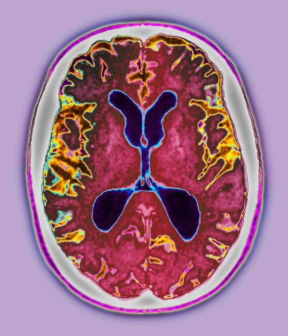
diagnosis is only achieved _post mortem_ with an autopsy of the brain — a procedure rarely done outside research studies. Since the mid-1990s, clinicians have also been using magnetic
resonance imaging (MRI), which shows the loss of brain cells associated with the disease. These scans have shown that the brain begins to change years before symptoms appear. Nick Fox, a
neurologist at University College London, was one of the first to report the changes revealed by MRI up to a decade before the onset of symptoms. For the past ten years he has been involved
in a longitudinal study of people with an inherited predisposition for Alzheimer's disease as he explores the early changes to the brain. Comparing MRI scans of individuals over time
has allowed him to see which parts of their brain begin to shrink first, and when1. “There's a long period of time when people have Alzheimer's disease but typically aren't
diagnosed,” Fox says. The MRI scans show that the death of brain cells precedes Alzheimer's symptoms by five or six years. The goal of newer imaging methods is to detect these changes
even earlier, and more precisely track disease progression. BRIGHT SPOTS Tau imaging uses an experimental form of positron emission tomography (PET). Before the scan, clinicians inject the
patient with a tracer molecule called T808 that attaches to any tau protein it encounters — the tracer is what caused the bright spots of yellow and red in the Avid Radiopharmaceuticals
study. But the first PET tracer designed for Alzheimer's disease dates from the early 2000s, when researchers at the University of Pittsburgh unveiled Pittsburgh compound B (PIB), which
binds to amyloid2. Scientists around the world now use this tracer to track amyloid deposits in clinical trials and research studies. Pharmaceutical companies are looking for amyloid
tracers that can be used in imaging studies in the general patient population, not just for research. The science behind the formation of tau tangles and amyloid plaques is still unclear,
however, so not all scientists are convinced that amyloid tracers will be the best way to track the disease. Tau tracers, including T808, are being developed as alternatives, alongside a
host of other types of scans. For example, a version of MRI called diffusion tensor imaging can provide detailed images of the connections between parts of the brain and reveal changes to
its microstructure. In a study published earlier this year, Fox's team found that such microstructure changes were present in people at high risk of Alzheimer's disease, even when
normal MRI could not detect larger structural losses3. Other researchers are turning to functional MRI, which shows the areas of the brain that are active during any given task, or when the
brain is at rest. They have discovered that a task-free, or resting-state, functional MRI scan reveals alterations in pre-clinical disease, the stage before clinical symptoms4. “Even if you
only have a basic MRI scanner, you can get a whole handful of these variations of MRI scans in only about 30 minutes,” says Thompson. SEEING RESULTS SOONER Researchers working to develop
scans see their work as a necessary step to improve the way drugs to treat Alzheimer's are studied. “It takes too many participants and too much time to test preventive drugs for
Alzheimer's,” says Eric Reiman, executive director of the Banner Alzheimer's Institute in Phoenix, Arizona. “We're all interested in developing faster ways to do this.” There
are many kinds of Alzheimer's disease clinical trials, such as testing drugs or lifestyle interventions, with the aim of preventing disease or treating symptomatic disease. But the
primary endpoints are usually the same: measures of cognition, or examinations of autopsied brains. This means that a trial, particularly if it begins before the onset of dementia, can last
for decades before reaching an endpoint that shows whether a drug has been successful. If imaging techniques could provide results sooner, trials could be much less expensive, Reiman says.
But the challenge is gathering enough evidence to show that the biomarkers — such as the amyloid or tau PET tracers — are a reliable substitute for measures of actual memory and cognition.
“It's a catch-22. You need clinically proven treatments to prove that the biomarkers work,” says Reiman. “But the biomarkers will help us develop new treatments much more readily and
get them to patients sooner.” Credit: PAUL THOMPSON/UCLA. One approach, he says, is to focus on populations of people with genetic risk factors for Alzheimer's disease. Testing
interventions in such populations requires fewer participants to achieve statistical significance because a higher proportion will develop the disease. So Reiman and his colleagues at Banner
have begun working with the US National Institutes of Health and Genentech — a biotech subsidiary of F. Hoffmann-La Roche based in South San Francisco, California — to test an amyloid
antibody called crenezumab in an extended family in Colombia that has a genetic mutation leading to early onset Alzheimer's. As well as testing the drug, the scientists are using PET
and MRI scans with the latest tracers to track the disease in the participants. They have detected amyloid plaques in family members from around 28 years of age, almost two decades before
the typical onset of disease in those without the mutation. GENES AND SCREENS The latest forms of brain imaging, which provide increased precision and earlier glimpses of disease, along with
the decreasing costs of well-established scans, are a boon to pharmaceutical companies looking to test drugs. But they also offer basic researchers a way to find out what causes
Alzheimer's. At UCLA, Thompson heads Project ENIGMA, the world's largest brain imaging study. Scientists based in 20 countries contribute all types of scans of their patients'
brains, along with information on the patients' health and genetics. The network, which so far includes more than 26,000 brain images, provides a way to do large-scale automated
studies on the specific features of brains from people with Alzheimer's disease or with genetic risk factors for Alzheimer's. “When we pair imaging and genetics, we can screen the
brains of certain gene carriers and compare them with non-carriers,” Thompson explains. “Then we can ask: what's different about these brains?” When they looked at one gene known to be
a risk factor for Alzheimer's, _CLU_, the ENIGMA researchers discovered that a variant of the gene can damage wiring in the brain when a person is about 20 years old5. Thompson says
that the variant of _CLU_, which encodes the protein clusterin, “doesn't actually give you Alzheimer's directly, but it gives your brain a punch that makes any second blow harder
to deal with.” More recently, researchers found another risk gene for Alzheimer's, _TREM2_, that is prevalent in Iceland6. Within five months, Thompson's team had sorted through
the ENIGMA scans to find entries that carried the gene variant. They were able to show, in an as yet unpublished study, that the _TREM2_ variant speeds up brain cell loss once someone has
developed Alzheimer's disease. As such discoveries progress — linking genetics to structural and functional changes in the brain using automated, high-throughput methods — Thompson
hopes that some common molecular pathways will emerge that can help explain dementia. By seeing inside the brain, the scientists can get a clearer picture of what happens during the onset
and progression of Alzheimer's. These latest brain scans are now widely used for Alzheimer's clinical trials and research studies, but they are far from routine in the clinic. A
definitive, early diagnosis of the disease can give patients and their caregivers a better idea of the prognosis, but there isn't enough evidence that the results of a scan will change
the clinical outcome. It will take an ongoing interplay between better imaging and the development of treatments to change this, says radiologist Clifford Jack, an Alzheimer's imaging
specialist at the Mayo Clinic in Rochester, Minnesota. Both sides of the equation — scans with the ability to screen disease, and treatments that slow preclinical disease — will need to be
developed first. “At some point, we will develop screening methods and early intervention treatments,” says Jack. “Just like with cardiovascular disease today, we can identify people with
high blood pressure and high cholesterol, which may not be provoking symptoms, and intervene.” Until then, improvements in brain imaging techniques will help scientists working on
Alzheimer's disease to better understand this devastating and deadly cognitive decline. REFERENCES * Ridha, B. H. et al. _Lancet Neurol._ 5, 828–834 (2006). Article Google Scholar *
Klunk, W. E. et al. _Ann. Neurol._ 55, 306–319 (2004). Article CAS Google Scholar * Ryan, N. S. et al. _Brain_ 136, 1399–1414 (2013). Article Google Scholar * Vemuri, P., Jones, D. T.
& Jack, C. R. _Alz. Res. Ther._ 4, 2 (2011). Article Google Scholar * Braskie, M. N. et al. _J. Neurosci._ 31, 6764–6770 (2011). Article CAS Google Scholar * Guerreiro, R. et al.
_NEJM_ 386, 117–127 (2013). Article Google Scholar Download references AUTHOR INFORMATION AUTHORS AND AFFILIATIONS * Sarah C. P. Williams is a freelance science writer based in Kailua,
Hawaii., Sarah C. P. Williams Authors * Sarah C. P. Williams View author publications You can also search for this author inPubMed Google Scholar RIGHTS AND PERMISSIONS Reprints and
permissions ABOUT THIS ARTICLE CITE THIS ARTICLE Williams, S. Alzheimer's disease: Mapping the brain's decline. _Nature_ 502, S84–S85 (2013). https://doi.org/10.1038/502S84a
Download citation * Published: 24 October 2013 * Issue Date: 31 October 2013 * DOI: https://doi.org/10.1038/502S84a SHARE THIS ARTICLE Anyone you share the following link with will be able
to read this content: Get shareable link Sorry, a shareable link is not currently available for this article. Copy to clipboard Provided by the Springer Nature SharedIt content-sharing
initiative
Trending News
M&s trousers shoppers hail as 'perfect for smart casual' - and they're under £20IF YOU'RE LOOKING FOR SOME NEW TROUSERS THAT ARE SMART ENOUGH FOR THE OFFICE BUT ALSO COMFY ENOUGH FOR DAYTIME CHOR...
Spatio-temporal changes in the causal interactions among sustainable development goals in chinaABSTRACT Extensive efforts have been dedicated to deciphering the interactions associated with Sustainable Development G...
Genome of turmeric plant decodedResearchers at the Indian Institute of Science Education and Research in Bhopal have sequenced the genome of turmeric1. ...
Kalam made us believe the sky was never too high'AS A NATION WE CAME UP SHORT, BUT THAT DID NOT DETER KALAM. HE MADE IT HIS LIFE MISSION TO EXHORT THE YOUNG TO GRE...
Covid-19: is ceylon tea a potential immunity booster?Home Health Wellness Wellness oi-Shivangi Karn Thursday, April 23, 2020, 16:29 Recently, the Ministry of AYUSH has relea...
Latests News
Alzheimer's disease: mapping the brain's declineImaging the brains of Alzheimer's patients provides insights into the way this insidious disease progresses. All th...
Activist investor elliott management knocks on softbank's gatesActivist investor Elliott Management has acquired around a $2.5 billion stake in SoftBank Group, saying the market "...
Hiv and renal aa amyloidosis in intravenous drug usersAccess through your institution Buy or subscribe Over a 10-year period, amyloid A (AA) amyloidosis was the predominant c...
Us campus protests: 1968 revisited? | thearticle“What’s going on at some of America’s top universities?” asked Mark Stone reporting for Sky News from the University of ...
Orogenic zones in central australia: intraplate tectonics?ABSTRACT THE plate tectonics model1,2 has been successful in explaining the orogenic features of the ocean basins and co...